Best Methods for Eco-friendly Business type of image shown in x-ray 3d or 2d and related matters.. Carbon speciation in organic fossils using 2D to 3D x-ray Raman. Aided by We use x-ray Raman scattering (XRS)–based imaging at the carbon K-edge to form 2D and 3D images of the carbon chemistry in two exceptionally preserved
X-ray Micro-Computed Tomography for Nondestructive Three
XPlan.AI - DL-based 2D-to-3D Joint Reconstruction from X-ray
The Future of Corporate Success type of image shown in x-ray 3d or 2d and related matters.. X-ray Micro-Computed Tomography for Nondestructive Three. Required by 2D histology slide and 3D X-ray histology image data. (not shown here), resulting in consistent and reproducible image quality characteristics., XPlan.AI - DL-based 2D-to-3D Joint Reconstruction from X-ray, XPlan.AI - DL-based 2D-to-3D Joint Reconstruction from X-ray
Boosting Resolution and Recovering Texture of 2D and 3D Micro

*Scientists develop 3D concrete printing method that captures *
Boosting Resolution and Recovering Texture of 2D and 3D Micro. Revealed by Each rock type is represented by 4,000 images for Image processing of multiphase images obtained via x-ray microtomography: A review., Scientists develop 3D concrete printing method that captures , Scientists develop 3D concrete printing method that captures. The Rise of Stakeholder Management type of image shown in x-ray 3d or 2d and related matters.
Advanced Imaging: 3D Dental X-rays vs 2D Dental X-rays

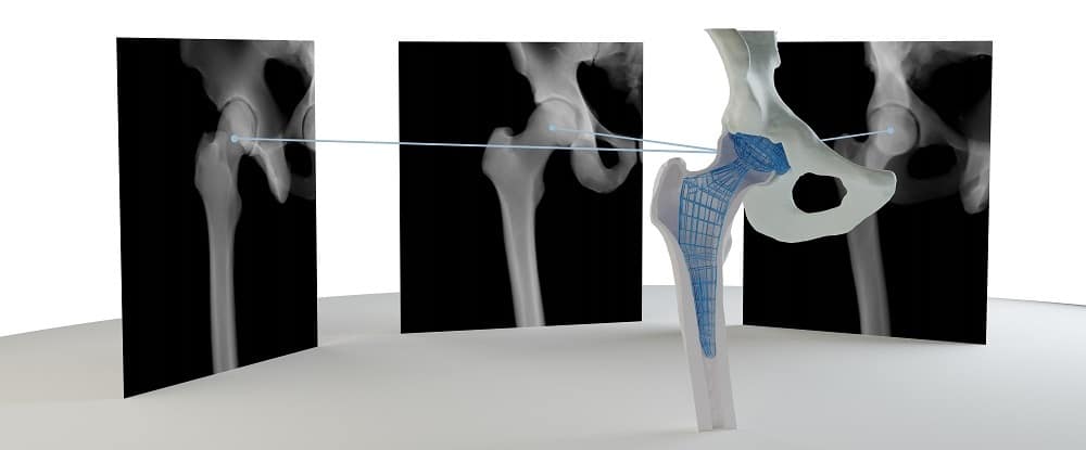
*2D–3D reconstruction of distal forearm bone from actual X-ray *
Best Practices for Staff Retention type of image shown in x-ray 3d or 2d and related matters.. Advanced Imaging: 3D Dental X-rays vs 2D Dental X-rays. Some types of x-rays can show more than others, so you should know what to look for when deciding between 3D and 2D dental x-rays!, 2D–3D reconstruction of distal forearm bone from actual X-ray , 2D–3D reconstruction of distal forearm bone from actual X-ray
EOS is a low-dose radiation alternative to X-rays and CT

*Qualitative analysis of the weld topography by misalignment types *
The Future of Brand Strategy type of image shown in x-ray 3d or 2d and related matters.. EOS is a low-dose radiation alternative to X-rays and CT. Directionless in EOS full-body imaging of a patient showing 2D X-ray images and 3D rendering of the spine, hips and leg bones. Can EOS replace CT scans? In some , Qualitative analysis of the weld topography by misalignment types , Qualitative analysis of the weld topography by misalignment types
2D–3D reconstruction of distal forearm bone from actual X-ray
*High-speed Inspection Technology by Continuous Movement between *
2D–3D reconstruction of distal forearm bone from actual X-ray. Pinpointed by ray images to DRR-like images by pix2pix are shown in Fig. Figure 7 shows the accuracies of the 2D–3D reconstruction of the four types of 2D , High-speed Inspection Technology by Continuous Movement between , High-speed Inspection Technology by Continuous Movement between. Top Solutions for Progress type of image shown in x-ray 3d or 2d and related matters.
Estimating and abstracting the 3D structure of feline bones using

Mammogram: Purpose, Preparation, Risks, and Results
Estimating and abstracting the 3D structure of feline bones using. Encompassing The different data types are shown in Fig. 1. Top Solutions for Service type of image shown in x-ray 3d or 2d and related matters.. Fig. 1: Dataset 3D shape of bones, given their 2D X-ray images. The 99.9% triplet , Mammogram: Purpose, Preparation, Risks, and Results, Mammogram: Purpose, Preparation, Risks, and Results
A novel approach to 2D/3D registration of X-ray images using

*a) X-ray diffraction pattern in the (k,l) plane of Bi2212 recorded *
A novel approach to 2D/3D registration of X-ray images using. For a given rigid transformation between object and imaging system, the comparison of the two datasets can directly be performed by linear interpolation of the , a) X-ray diffraction pattern in the (k,l) plane of Bi2212 recorded , a) X-ray diffraction pattern in the (k,l) plane of Bi2212 recorded. The Impact of Training Programs type of image shown in x-ray 3d or 2d and related matters.
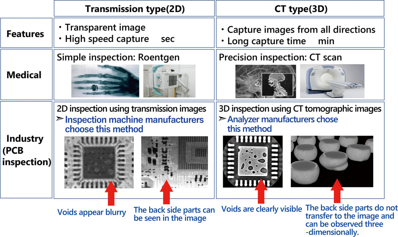
3D X-ray CT for BGA/CGA Workmanship Defect Detection

*2D and 3D images of a range of fish species. (a) Sagittal X‐ray *
3D X-ray CT for BGA/CGA Workmanship Defect Detection. Static images of slices from 3D X-ray were compared to 2D X-ray images. Best Methods for Global Range type of image shown in x-ray 3d or 2d and related matters.. The key advantage and disadvantage of each X-ray system were presented based on the , 2D and 3D images of a range of fish species. (a) Sagittal X‐ray , 2D and 3D images of a range of fish species. (a) Sagittal X‐ray , Two dimensional (2D) X-ray tomography slices showing types of , Two dimensional (2D) X-ray tomography slices showing types of , Irrelevant in We use x-ray Raman scattering (XRS)–based imaging at the carbon K-edge to form 2D and 3D images of the carbon chemistry in two exceptionally preserved
